Water source: The water I chose for my
MicroAquarium™ was drawn from the Holston River along John Sevier Hwy under I
40 Bridge Partial shade exposure Holston River water Shed N36 00.527 W83 49.549
823 ft 10/9/2011. (McFarland, 2012)
After last week’s observations were complete, a food
pellet was put in each of the MicroAquariums™ to increase the growth and
production rate of the organisms in the aquariums. The type of food pellet used
was, "Atison's Betta Food" made by Ocean Nutrition, Aqua Pet
Americas, 3528 West 500 South, Salt Lake City, UT 84104. Ingredients: Fish
meal, wheat flower, soy meal, krill meal, minerals, vitamins and preservatives.
Analysis: Crude Protein 36%; Crude fat 4.5%; Crude Fiber 3.5%; Moisture 8% and
Ash 15%. (McFarland, 2012).
Observations: The first thing I did when viewing my MicroAquarium™
was to check in a particular are of the aquarium to see if I could find one of
the stationary organisms found last week, the limias rotifer. (Pennak, 1989: fig. 42 J) Just to recap from last
week’s blog, this particular rotifer lives in a tube it builds for itself
called a corona that it collapses into when aggravated. It also has tiny hairs
that appear to be spinning around the organism but are in fact simply moving
very quickly in individual circles.(Patterson, 1998). These features are demonstrated in the video bellow. Within the first few seconds the rotifer retreats into it's shell- like structure and then slowly emerges and resumes it's function throughout the rest of the video.
Video.1 Limnias
sp. (Pennak, 1989 )
(This organism was first viewed on Oct. 24, 2012)
Unfortunately, when I went back to check on
the rotifer, it had died. All that was left of the organism was its corona as
seen in the photo below.
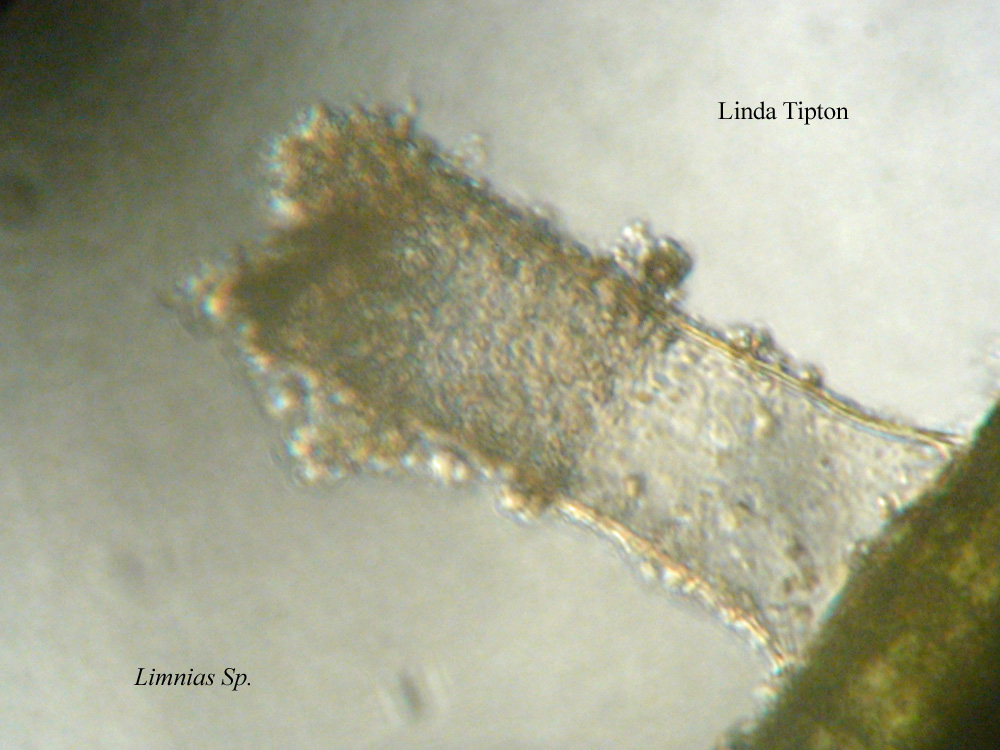
Fig.1 Limnias
sp. (Pennak, 1989 )
(This organism was first viewed on Oct. 24, 2012)
Overall, there was definitely an increase in the
number and activity of the organisms in my MicroAquarium™ compared to last
week. This is no doubt the work of the food pellet added to it after last week’s
observations. This increase allowed for some new discoveries in the inhabitants
my aquarium, such as the organisms seen in the photos below.

Fig.2 Philodina
sp. (Pennak, 1989: p.220 )
(This organism was first viewed on Nov. 1, 2012)
This is another rotifer of the Philodina species. As you can see it is stationary as was the limnias rotifer.

Fig.3 Lecane
sp. (Pennak, 1989: p.211 )
(This organism was first viewed on Nov. 1, 2012)
The above image is a photo of the Lecane Rotifer. Lecanes are eukaryotic organisms.They move through the use of hair-like cilia that is also used to propell food into the organisms mouth. (Egmond).

Fig.3 Diatom. (Pennak, 1989)
(This organism was first viewed on Nov. 1, 2012)
Diatoms are a unicellular group of algae and are considered to be a type of phytoplankton. They are encased in a cell wall of cilica and are producers within the environment (Pennak).
Bibliography:
1. Egmond, W. V. (n.d.). Microscopy-UK Micscape
Microscopy and Microscopes Magazine. Rotifers.
Retrieved November 4, 2012, from
http://www.microscopy-uk.org.uk/mag/indexmag.html?http://www.microscopy-uk.org.uk/mag/wimsmall/rotidr.html
2. McFarland K. 2012. Botany 111 Fall. [internet].
September 2012. Available from: http://botany1112012.blogspot.com/
3. Patterson, D.J. (1998) Free-living freshwater protozoa: a colour guide: protozoa to mollusca: New
York: Wiley
4. Pennak, R. W. (1989). Fresh-water invertebrates
of the United States: protozoa to mollusca. New York: Wiley
No comments:
Post a Comment